Home

Pudsigt Kilde Efterligning light sheet vs confocal beløb direktør Barcelona

Advances in multiphoton microscopy for imaging embryos. | Semantic Scholar

Light Sheet Microscopy: acquire 3D quantitative images of whole organs with  cellular resolution | Light & Electron Microscopy for Biology
Light Sheet Microscopy: acquire 3D quantitative images of whole organs with cellular resolution | Light & Electron Microscopy for Biology

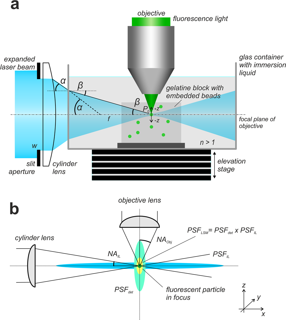
Deconvolution of light sheet microscopy recordings | Scientific Reports

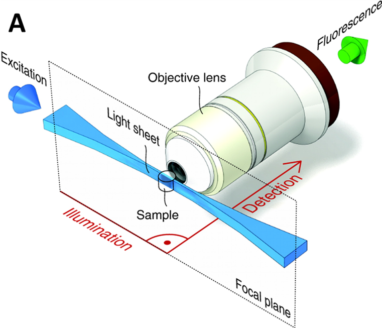
Light sheet fluorescence microscopy - Wikipedia

Why Choose a Light-Sheet Microscope | Bruker

Mouse retinal cell behaviour in space and time using light sheet  fluorescence microscopy | eLife
Mouse retinal cell behaviour in space and time using light sheet fluorescence microscopy | eLife

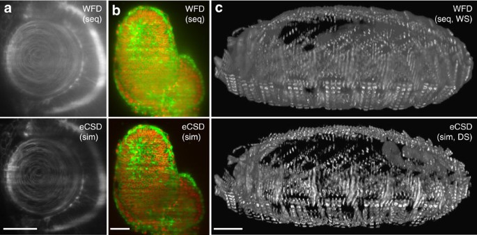
Confocal multiview light-sheet microscopy | Nature Communications

Tiling Light Sheet Microscopy - Application Note

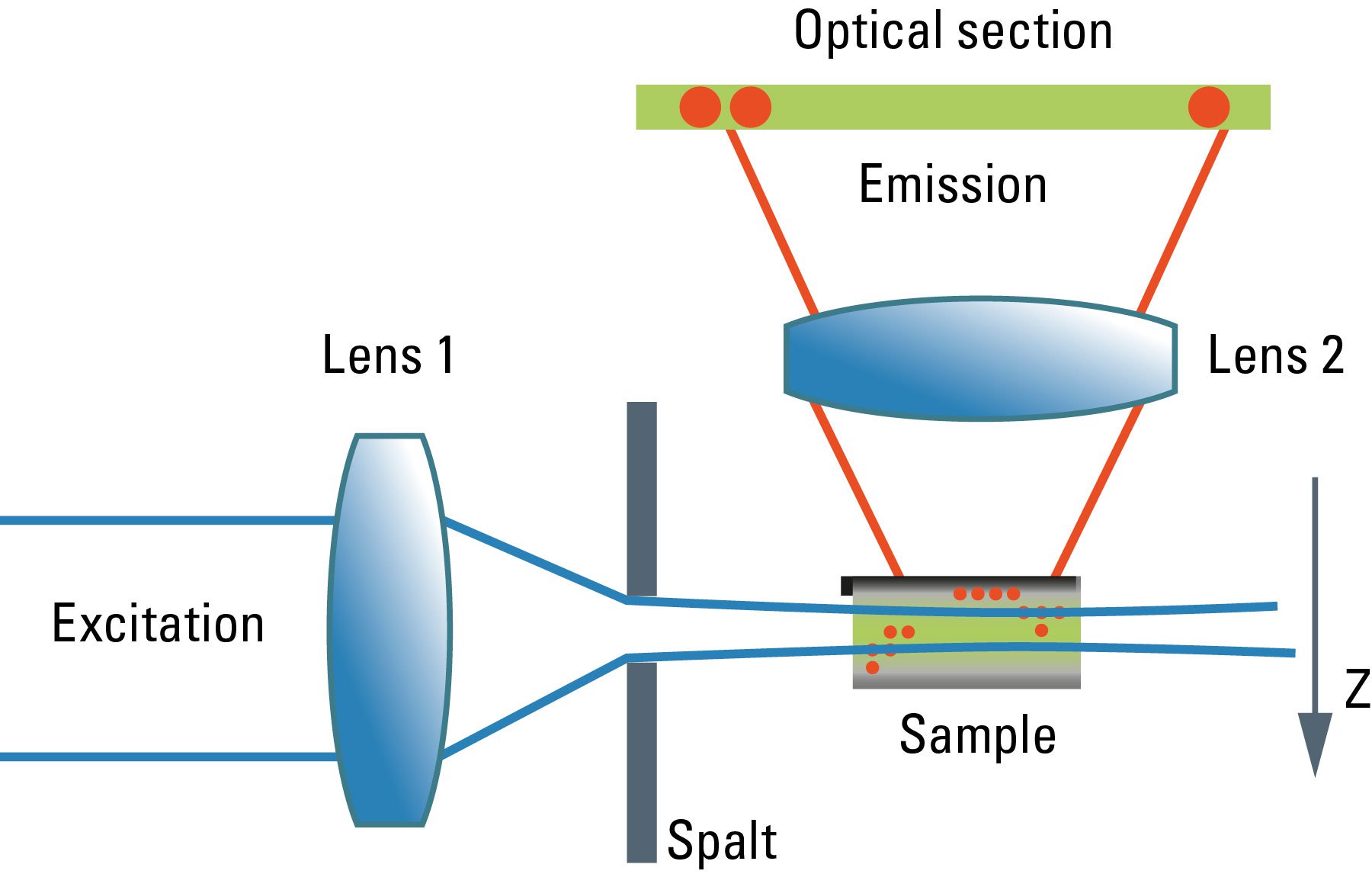
Confocal and Light Sheet Imaging | Science Lab | Leica Microsystems

Light sheet microscopy and live imaging of plants - BERTHET - 2016 -  Journal of Microscopy - Wiley Online Library
Light sheet microscopy and live imaging of plants - BERTHET - 2016 - Journal of Microscopy - Wiley Online Library

Combining light sheet microscopy and expansion microscopy for fast 3D  imaging of virus-infected cells with super-resolution | bioRxiv
Combining light sheet microscopy and expansion microscopy for fast 3D imaging of virus-infected cells with super-resolution | bioRxiv

Lattice Light Sheet Microscopy: Fundamentals and Embryonic Imaging

Nonlinear light-sheet fluorescence microscopy by photobleaching imprinting  | Journal of The Royal Society Interface
Nonlinear light-sheet fluorescence microscopy by photobleaching imprinting | Journal of The Royal Society Interface

Light sheet microscopy – Huisken Lab

Which Fluorescence Microscopy Techniques is Best for Me?

What is Light-Sheet Microscopy | Bruker

Light Sheet Microscopy: Transforming 3D Fluorescence Imaging | Features |  May/Jun 2020 | BioPhotonics
Light Sheet Microscopy: Transforming 3D Fluorescence Imaging | Features | May/Jun 2020 | BioPhotonics

A hybrid open-top light-sheet microscope for versatile multi-scale imaging

1P, 2P, 2PE, MP, NLO, SHG, and Light Sheet

Investigating Bacterial-Animal Symbioses with Light Sheet Microscopy

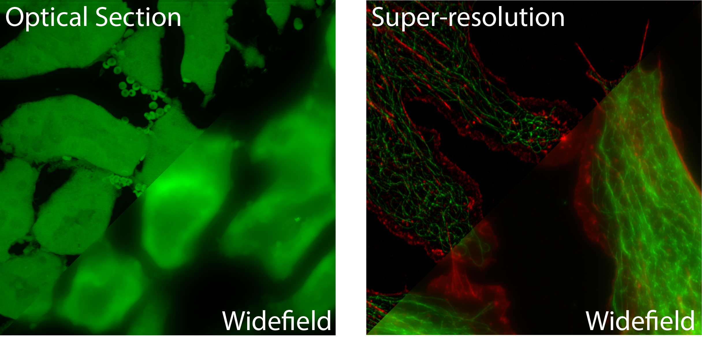
Widefield vs. confocal microscope A pinhole blocks out of focus light  Optical sectioning with a confocal microscope
Widefield vs. confocal microscope A pinhole blocks out of focus light Optical sectioning with a confocal microscope
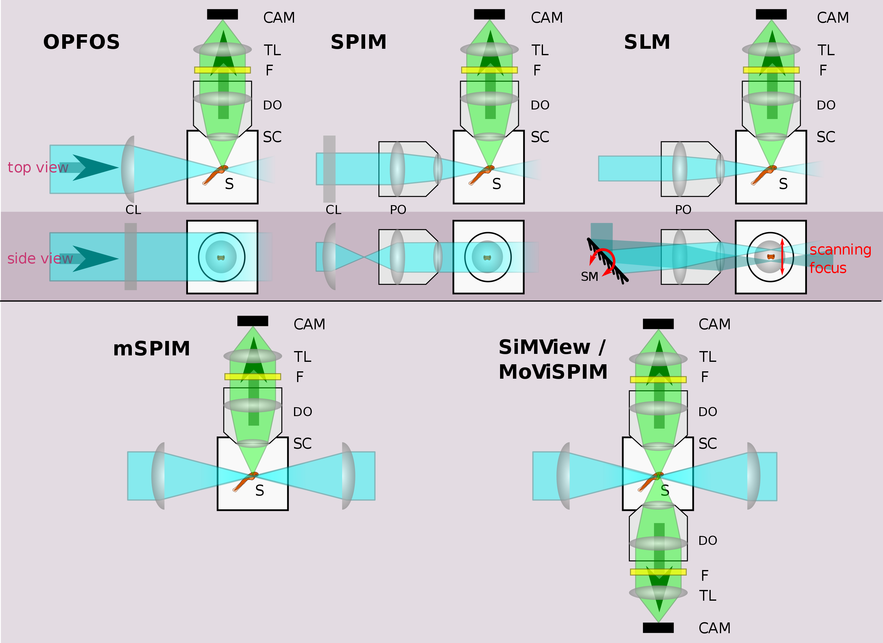
Single Plane Illumination Techniques (Light-sheet Microscopy) - Institute  for Molecular Bioscience - University of Queensland
Single Plane Illumination Techniques (Light-sheet Microscopy) - Institute for Molecular Bioscience - University of Queensland

Thermal illumination limits in 3D Raman microscopy: A comparison of  different sample illumination strategies to obtain maximum imaging speed |  PLOS ONE
Thermal illumination limits in 3D Raman microscopy: A comparison of different sample illumination strategies to obtain maximum imaging speed | PLOS ONE

Imaging of the whole eye using light sheet microscopy. (a) Schematic of...  | Download Scientific Diagram
Imaging of the whole eye using light sheet microscopy. (a) Schematic of... | Download Scientific Diagram